Confirmed placement af-ter reading the x-ray. Tube feedings were initi-ated.

Learningradiology Dobhoff Dobbhoff Tube Malplaced Rll
Dobhoff Tube Placement.

. At our facility we x-ray all feeding tubes for placement verification. What is a Doboff Tube Placement procedure. Nasogastric NG tube position on chest x-ray should be assessed following initial placement and on subsequent radiographs.
Abdominal x-ray after fluoroscopic guided Dobhoff tube placement. X-rays are only performed when the position is uncertain. Steps for NG Feeding Tube Placement in an Awake Patient.
On HD 5 the patient underwent a fluoroscopic guided Dobhoff tube placement to maintain enteral nutrition. Aspiration and a Dobhoff tube was subsequently placed at the bedside for delivery of enteral nutrition. The tube is inserted into the stomach by way of the nasal passage.
The tube is inserted by the use of a guide wire called the stylet see image1 which removed after the tube correct placement is confirmed. Abdominal X-ray after Dobhoff tube DHT placement to confirm accurate positioning. Nasoduodenal ND feeding tube placement is a procedure in which an x-ray monitor is used to guide the placement of a soft feeding tube through the nose into the small bowel duodenum.
Tube feedings were begun. The patient experi-enced respiratory dis-tress. A Dobhoff tube has a weighted end that helps guide it through.
120cm preferred over blue tip dobhoff tube Lubricant 60 ml syringe. Follow-up abdominal x-ray revealed displacement of the Dobhoff tube in the left pleural space. ND feeding tubes may be used for long-term enteral nutrition.
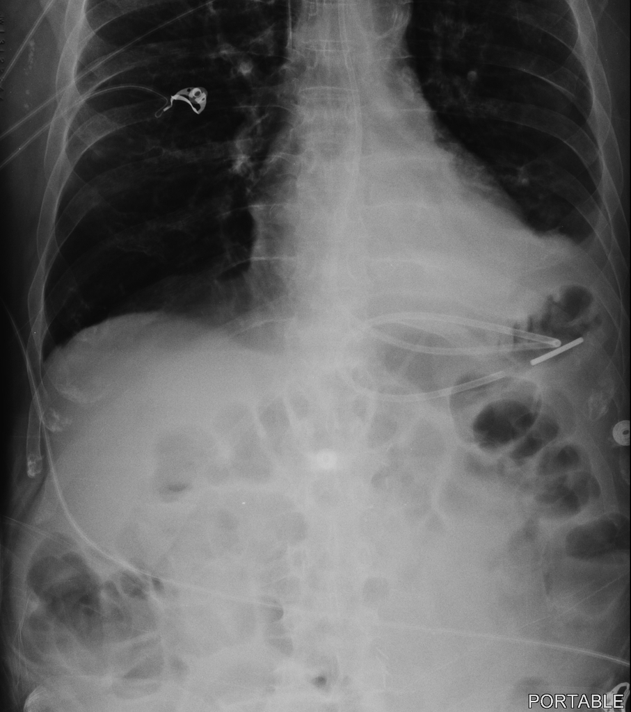
2 What you need zINFORMED CONSENT zNG or dobhoff tube zLubricant z60cc syringe zCup of water and straw zStethoscope Tube placement Ideally patient should be in sniffing position neck flexed head extended Also in a perfect world. B The tip of the Dobbhoff tube is in the descending limb of the. Auscultation was performed in all 78.
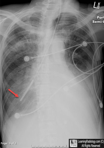
Microsoft PowerPoint - NG Feeding Tube GME Education_Medicinepptx Author. A Spray nasal passage with oxymetazoline b Anesthetize nasal passage and oropharynx with lidocaine or benzocaine Measure how. A review of the x-ray showed that the feeding tube was in the main bronchus.
This tube is necessary to give the patient tube feedings. A The tip of the feeding tube white arrow is in the right lower lobe bronchus having descended in the trachea instead of the esophagus. Thus DHT insertion requires radiologist confirmation of correct placement with chest x-ray CXR increasing clinical delays.
The patient was found dead. A Dobhoff tube can be inserted at a patients bedside by a nurse or physician. Of 78 nasoenteral intubations in 46 patients using a Dobbhoff Biosearch Medical Products weighted enteral feeding tube gastric aspirates were evaluated in 28.
Gastrointestinal anatomy before and after Roux-en-Y. The stylet is removed after the tubes correct placement has been confirmed. In order to prepare a patient for the insertion of a Dobhoff tube the esophagus and nasal cavity are numbed and the patient if conscious may be given a mild sedative.
Patients are usually positioned on the right side while the tube is put into the nose. We have a more in-depth reference article NGT. Blind Placement with radiographic confirmation Blind technique is defined as the clinician relying on manual feel.
The tube is inserted by the use of a guide wire called the stylet see image1 which removed after the tube correct placement is confirmed. Chest x-ray revealing bilateral chest tubes see arrows with near resolution of bilateral pneumothorax. The tube was actually named after two physicians Drs.
The Dobhoff tube was introduced in the mid-1970s by surgeons. Small amount of contrast injected to confirm Dobhoff tube see arrow positioning in the fourth portion of the duodenum. Dobhoff tube is a special type of nasogastric tube NGT which is a small-bore and flexible so it is more comfortable for the patient than the usual NGT.
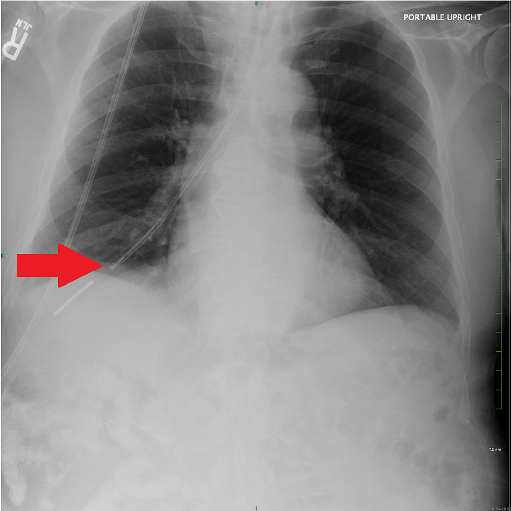
Dobhoff tube is an excellent alternative although it can carry a serious risk of pneumothorax if placed blindly. A chest X-ray performed shortly after the tube placement demonstrated that the tip of the Dobhoff tube was within the right lung base following the course of the right mainstem bronchus Figure 1. Follow-up abdominal x-ray revealed displacement of the Dobhoff tube in the left pleural space.
Posted Nov 18 2004. Fluoroscopy-Dobhoff Tube Placement. We had a situation where a dubhoff did not show up anywhere on the x-ray.
After removal of the tube a follow-up chest x-ray revealed iatrogenic bilateral pneumothoraces. During each tube insertion the colorimetric CO 2 detector was used and the color change was noted which is representative within the results section. Tracheobronchial insertion of DHTs presents a significant risk for pulmonary complications.
The x-ray was read and placement confirmed. Four Different Patients with Dobbhoff feeding Tubes. Dobhoff tubes come with a radiopaque stripe them easily visible by x-ray.
The distal tip of the feeding tube is in a loop of jejunum in patient status post gastrojejunostomy. Proper Dobhoff Placement PICO Question In Adult ICU patients will a two-step placement protocol improve RN small bore feeding tube insertion competence compared to LVHN standard practice policy. Therefore bilateral chest tubes were placed.
A Dobhoff tube was placed by a house physi-cian. A guide wire called a stylet is used during insertion. Acute hypoxemic respiratory failure ensued.
The Dobhoff tube was advanced through the right nasal cavity and the tube was confirmed to be in the distal esophagus. Patients 8 patients had numerous x-ray images due to multiple Dobhoff NG tube insertions. When a Cortrak machine is not available a two step technique using a chest x-ray to confirm placement in the esophagus and then through the pylorus is an effective way to prevent serious complications23 rEFErEncES 1.
Nov 18 Okay heres a question Dobhoff tube placement. Dobhoff tubes DHT are narrow-bore flexible devices that deliver enteral nutrition for critically ill patients. This is a summary article.
After removal of the tube a follow-up chest x-ray revealed iatrogenic bilateral pneumothoraces. An x-ray can ensure that the Dobhoff tube has been placed correctly. The wire was left in.
Data was collected at initial placement prior to x-ray confirmation. I have a question for anyone who can help me. It was a 12 french by the way.
Dobhoff tube is a special type of nasogastric tube NGT which is a small-bore and flexible so it is more comfortable for the patient than the usual NGT. After recently an update to our Policy and Procedure for gastric tube placement I noticed a statement which instructed the RN to leave the guidewire in-place until after radiographical confirmation was obtained. Always check the final Xray reading before beginning tube feeds.
One patient had 10 Dobhoff NG tube insertion attempts which resulted in 10 x-ray images. Dobhoff tube placement is performed when a patient is unable to drink or eat due to various reasons.

Abdominal X Ray Revealing The Dobhoff Tube Traversing The Left Main Download Scientific Diagram

Southwest Journal Of Pulmonary Critical Care And Sleep Imaging Medical Image Of The Week Dobhoff Tube Placement With Roux En Y Gastric Bypass
Learningradiology Dobhoff Dobbhoff Tube Malplaced Rll

Iatrogenic Bronchopleural Fistula From A Dobhoff Tube Radiology Case Radiopaedia Org
Icu Chest Radiography Lines Ng Dobhoff Etc Youtube
Cureus Hemothorax Following Traumatic Dobhoff Tube Insertion
2
Dobhoff Nasogastric Tube Tube Radiology Case Radiopaedia Org
0 comments
Post a Comment